Venipuncture is the process of obtaining blood samples from veins for the purpose of laboratory testing.
The procedure is performed by phlebotomists, paramedics and medical laboratory scientists, to name a few.
Venipuncture is probably the most common procedure in the medical field; usually performed for either of the following reasons:
- To obtain blood samples in order to perform diagnostics
- To administer therapeutic treatments to patients
- To collect blood for later use should the patient’s condition requires transfusions
- To remove blood that was found with excessive levels of erythrocytes or iron
- To monitor the levels of various blood components
- Patient Relations And Identification
- Patient’s Bill of Rights
- Infection Control And Safety
- Equipment Necessary to Perform a Routine Venipuncture
- Vein Selection
- The Venipuncture Procedure
- Additional Considerations in Relation to Venipuncture (Preventing Hemolysis, Hematoma and Negative Effects of Prolonged Tourniquet Use)
- Special Considerations (Muscular activity, exercise effects, stress)
- Therapeutic Drug Monitoring
- Diurnal Rhythms
- Postural Changes
- Troubleshooting an Incomplete Collection/No Blood Obtained
- No Blood Collection
- Other Issues
- Order of Draw for Vacutainer/Microtainer Tubes
- External References
Patient Relations & Identification
As a phlebotomist, you must be courteous, professional and understanding in all your contacts with patients. The first thing you need to do is greet the patient and let them know whom you are and what you are going to do. Remember that effective communication is not only verbal, but nonverbal as well.
It is MANDATORY that you properly identify every patient prior to drawing his blood. If a patient is alert, ask him to tell you his full name.
NEVER say his name for him and just ask for a yes or no. You also need to check the patient’s ID bracelet to ensure this patient is who he says he is.
NEVER draw blood from an inpatient that does not have an ID bracelet. If you are working with inpatients, a member of the nursing staff should be able to help you by identifying the patient for you.
An outpatient is required to provide you with another form of identification as well. For each outpatient, you will be given a requisition. You must ask the patient for his birth date and last name to ensure this is the patient on your requisition.
Should there be any identification issues, a government issued photo ID card will suffice.
During the venipuncture procedure, try to take the patient’s mind off of the process by speaking with him. A patient who feels at ease will focus less upon the procedure. Once you have completed your task, always remember to thank the patient and courteously excuse yourself.
Patient’s Bill of Rights
Many hospitals have adopted the Joint Commission on Accreditation of Healthcare Organization’s (JCAH) Patient’s Bill of Rights. The bill of rights that are endorsed by the JCAHO are given below (in condensed form).
Each patient is entitled to:
- Impartial access to accommodations or treatment that is available or medically indicated despite his race, sex, creed, source of payment for his care or national origin.
- Respectful and considerate care that includes confidentiality of all records or any other communications pertaining to his care. Expect that all information including consultation or discussion about each patient’s case be conducted discretely. Individuals who are not directly involved in a patient’s case should not be present without the explicit consent of the patient.
- Know the professional status and identity of individuals who are providing his services. He must also know which practitioner or physician is primarily responsible for his care.
- Expect reasonable safety that is congruent with the practices of the hospital and environment.
- Practical informed participation in the decisions involving his healthcare. He shall be informed if the hospital plans on engaging in or performing research/education or human experimentation that will affect his treatment or care. Patients have the right to refuse to participate in such activities.
- Receive from his practitioner all the details related to his treatment, diagnosis and prognosis. This information must be given to the patient in terms that he can reasonably be expected to understand.
- Refuse obtaining treatment (to the extent that is permitted by law).
- Consult with a specialist at his request and expense.
- Be fully informed of the hospital’s rules and regulations in regards to patient conduct.
- Despite the payment source, he has the right to request and receive a detailed, itemized bill that explains the services the hospital rendered him.
Infection Control & Safety
Phlebotomists are exposed to sick patients and hazardous specimens. Following all safety and infection control procedures is essential.
Keep universal precautions in the forefront:
- Always wear a lab coat/gown and clean gloves when handling any body fluids.
- Frequently wash your hands.
- Change your gloves following contamination and with each patient.
- Dispose of all biohazard materials in appropriate containers.
- Clean up blood spills using a disinfectant solution that contains 10 percent bleach.
- Always dispose of contaminated needles immediately after removing it from the patient’s vein. Never recap, break, re-sheath or bend needles. This avoids splashing of its contents and accidental needle punctures.
- Place all your blood collection equipment away from the patients. This is especially true for children and psychiatric or geriatric patients.
Equipment Necessary to Perform a Routine Venipuncture
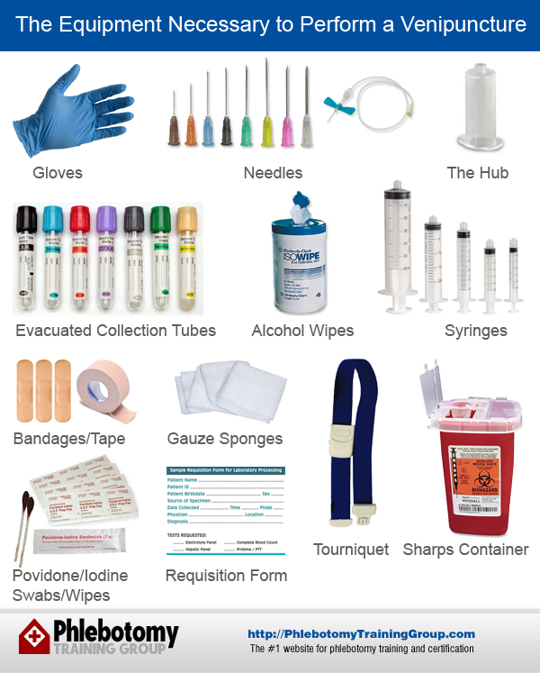
Gloves
These gloves can be constructed of rubber, latex, vinyl, etc. You will wear these to protect yourself and the patient.
Needles
Each needle has a gauge number. This number indicates the size of the needle’s bore. The larger the number, the smaller the needle’s bore is. There are needles that are used with evacuated blood collection systems and for use with a syringe, butterfly system or a single draw. For a detailed needle gauge comparison, click here.
Evacuated Collection Tubes
These tubes have a vacuum inside that is designed to obtain a predetermined volume of blood. There are various colored rubber stoppers on these tubes. The color of the stopper indicates which additive the tube contains.
Never pour blood from one tube into another. The tubes could have different coatings or additives in them.
The Hub
This holder/adapter is used with the evacuated tube collection system.
Alcohol Wipes
Isopropyl alcohol (70 percent) to sanitize the venipuncture site.
A Tourniquet
This is used to make feeling a vein easier for you.
Gauze Sponges
These are applied to the site following the removal of the needle.
Povidone/Iodine Swabs/Wipes
These are used to draw blood cultures and test alcohol levels in the blood.
Adhesive Tape/Bandages
These protect the venipuncture site following the blood collection procedure.
Syringes
A syringe is sometimes used with a butterfly needle instead of the evacuated collection system for special circumstances. Special circumstances include drawing from a patient’s hand or a child’s small vein.
Needle Disposal Unit/Sharps Container
Needles should NEVER be recapped, broken, bent as this could be extremely dangerous. Needles must be disposed of immediately following the withdrawal. Needles must be placed in a disposal unit that has been specifically designed for needle disposal.
Requisition/Order Form/Labels
A requisition/order form or labels are required for each sample that is submitted to the laboratory. Each form/label has to contain all the information necessary to process the specimen.
Essential Elements on the Requisition
- The patient’s full name.
- The patient’s sex and date of birth.
- The patient’s ID number.
- The test the physician is ordering.
- The full name of the physician ordering the test.
- The date and time of collection.
- The collecting phlebotomist’s initials.
- The source of the specimen. This information is necessary when a physician is requesting a fluid analysis, microbiology, cytology or any other testing where the analysis and reporting is site specific.

A sample Requisition Form. Based on information from the University of Utah.
Vein Selection
 While the fuller, larger cephalic and median cubital veins are drawn from the most frequently, the basilica vein that is located on the dorsum of the arm and dorsal hand veins are also acceptable sites for venipuncture. Due to the high probability of complications, foot veins are only used as a last resort.
If you cannot see superficial veins, you can bring the blood into the vein if you massage the patient’s arm starting at his wrist and moving to his elbow and/or lowering the patient’s arm by placing it beside the bed/chair.
If the first two techniques do not accomplish the task of bringing blood to the vein you could try placing a warm, damp washcloth on the site for approximately five minutes, gently pat the site or tapping the site using your thumb and index or thumb and second finger.
Although visually looking for a vein can assist you in locating one, you should never perform a venipuncture using sight alone. You need to palpitate and then trace the path of the patient’s veins using your finger.
Most phlebotomists begin their career using their index finger for this task. However, you may find that your second or ring finger is more sensitive for this task. If this is the case, then use that finger instead.
Knowing the difference between an artery and a vein is simple. An artery will pulsate and is more elastic than a vein. If a vein is thrombosed, it will lack resilience and feel like a cord, this kind of vein also rolls easily.
Avoid performing a venipuncture on:
- The upper arm of a patient who has had a mastectomy on that side. If drawn here, the test results could be inaccurate because of lymph edema.
- Sites that have extensive scarring from surgery or burns. Performing a venipuncture at these sites is more difficult due to the scar tissue.
- A site that is on the same arm as an IV/blood transfusion. The fluid in the IV could dilute the specimen. If avoiding the IV/blood transfusion arm cannot be helped, a procedure can be followed.
- A hematoma could cause incorrect test results. If there is not another site available, draw distal to the hematoma.
- Edematous extremities should be avoided because the accumulated fluid could alter test results.
- Arms that have heparin locks/fistulas/cannulas should not be drawn from without the permission of the patient’s physician.
The Venipuncture Procedure
It requires skill and knowledge to properly perform a venipuncture. A phlebotomist will generally establish her own routine.
- Approach each patient in a friendly, calm manner. Gain a patient’s cooperation by providing him with as much comfort as possible during the procedure.
- Ask the patient his name and date of birth to ensure proper identification. Always check his ID bracelet/ID prior to drawing his blood.
- Some patients have allergies to adhesives, antiseptics and/or latex. Many times inpatients will be wearing an armband stating this fact. However, you should make it a habit to ask each patient if he has an allergy to any of the products you will be using.
- Evaluate the patient’s disposition. This means that if any of the tests ordered by the physician require the patient be fasting, you need to find out when the last time was that he ate or drank anything besides water. Technological advancements have provided us with the ability to print labels that can be attached to each tube. However, there may be times when, for whatever reason, you must fill out a requisition form and indicate which tests a physician has ordered. Because of this possibility, you need to know how to accomplish this task.
- Scan the requisition form/labels for the patient’s information, the tests that are ordered and any other special requirements. The patient should be sitting, lying down or sitting up in his bed.
- Put your gloves on.
- Hyperextend his arm.
- Select an appropriate venipuncture site.
- Prepare your equipment, the patient and the chosen site.
- Properly place the tourniquet around the patient’s arm about 3 or 4 inches higher than the puncture site you have selected. Remember not to place the tourniquet on tightly or keep it on the patient’s arm any longer than 1 minute to avoid hemoconcentration of the blood sample. If you remove the tourniquet, you must wait 2 minutes before you can apply it again.
- Ask the patient to make a fist (without pumping his hand).
- Now, select the vein you intend to draw from.
- Prepare his arm using the alcohol prep. You should use a circular motion to clean the site, starting inside and then working out. Allow the site to dry. If you are drawing blood cultures or blood alcohol levels, you need to use the povidone/iodine swabs/wipes instead.
- Hold the patient’s arm firmly and use your thumb to anchor the vein. Insert the needle swiftly at a 15 to 30 degree angle. Avoid excessive probing.
- Perform the venipuncture procedure collecting the sample in the correct collection tubes using the correct order of draw.
- While the final tube fills, remove the tourniquet.
- Gently place the gauze over the puncture site and swiftly withdrawal the needle using a backward motion.
- Once you have fully removed the needle, place it in the sharps container while pushing down on the gauze. Adequate pressure should keep a hematoma from forming.
- Dispose of any other contaminated supplies in the designated biohazard containers.
- If a tube has an additive, mix it by gently rocking it back and forth eight times.
- Appropriately label all the tubes while you are still with the patient.
- Remove your gloves and dispose of them in a biohazard container. Wash your hands.
- Deliver the labeled specimens to the laboratory in a prompt, efficient manner.
Proper Procedure for Drawing Beneath an IV
- Ask the patient’s nurse to turn off the IV for about two minutes prior to the venipuncture procedure.
- Once the two minutes have passed, apply the tourniquet beneath the IV site.
- Choose a vein, other than the vein with the IV.
Now you can perform the venipuncture. You need to draw 5 ml of blood and then discard it prior to drawing the blood for testing.
While drawing blood from and intravenous line could avoid venipuncture, there are other problems introduced. The line being drawn from must be flushed and if the blood is withdrawn too quickly, hemolysis could occur.
The Equipment Necessary to Perform a Fingerstick or Heel Stick
- Gloves.
- Physician’s orders/labels or requisition.
- Isopropyl alcohol wipes (70 percent) to sanitize the puncture site.
- Gauze sponges.
- A sharps container.
- Microtainer collection tubes, a glucometer, neonatal screen and/or capillary tubes.
- A warm washcloth (depending).

Performing a Fingerstick
- Put on your gloves. The ideal locations for a fingerstick are the middle or ring finger of a patient’s non– dominant hand.
- Sanitize the puncture site with the alcohol wipe in a circular motion. Allow the area to air dry.
- Use a sterile lancet to make a skin puncture next to the center of the finger pad, puncture perpendicular to the fingertip’s ridges. This ensures that the drop of blood does not run down the ridges of the fingerprint.
- Dispose of the lancet in your sharps container.
- Always wipe away the initial drop of blood with the gauze. This drop tends to have excess tissue fluid in it.
- Collect the drops of blood into the proper collection device. You can gently massage the finger; however, avoid excess pressure.
- Cap and rotate the collection device back and forth to mix the blood if there are any additives in the tube.
- Ask the patient to hold gauze over the puncture site for a few minutes until the bleeding subsides.
- Dispose of the contaminated materials in the designated containers.
- Properly label the tubes while still with the patient.
- Check the patient’s puncture site to ensure the bleeding has ceased.
- Remove your gloves and dispose of them properly. Wash your hands.
- Deliver the specimens to the lab.
Areas of Avoid Performing a Fingerstick
The center of and the tip of the finger should be avoided. The side of the finger has less soft tissue and this is where the nerves and vessels are located, the bone is also closer to the surface here.
The fifth finger does not usually have very much soft tissue over the bone.
Do not puncture a finger that is swollen, cold, covered with a rash or scarred.
The index finger usually has thicker, callused skin and may be more difficult to puncture.
Performing a Heel stick
- Follow all the greeting and preparation procedures as listed above. Heel sticks are recommended to obtain blood from newborns or infants.
- Pre–warming the heel (for 3 to 5 minutes) is recommended. Be sure you do not use a hot temperature; babies have thin skin that is extremely susceptible to thermal injury. Hold the baby’s foot firmly during collection. Never puncture the heel in the central portion. The bone is close to the surface there. Use the side areas as directed in class.
- Wash your hands with the recommended washing procedures for use in the nursery.
- Dry your hands.
- Put on your gloves.
- Sanitize the puncture site.
- Use a sterile lancet to cut across the heel’s print lines. This helps the blood well up for easier collection.
- Use clean gauze to wipe the first drop of blood away.
- Use a very gentle pressure to assist the blood flow.
- Fill the microtainer, capillary tubes or neonatal screen, as ordered.
- Once you have finished the procedure place gauze on the puncture site and elevate the baby’s heel.
- Dispose of the lancet in the sharps container and dispose of contaminated materials in the proper waste receptacles.
- Label the collection tubes, neonatal screen or capillary tubes appropriately.
- Remove the gloves.
- Wash your hands.
- Deliver the specimens to the lab.
Proper Safety Procedure to Follow If You Stick Yourself with a Contaminated Lancet or Needle During a Venipuncture
- Promptly remove and properly dispose of your gloves.
- Squeeze the puncture site as this promotes bleeding.
- Wash this area with soap and water.
- Note the patient’s ID number and name.
- Follow the guidelines as set forth by the institution you work at regarding treatment and follow–up procedures.
Using prophylactic zidovudine following exposure to blood contaminated with HIV is 79 percent effective in preventing seroconversion.
Additional Considerations in Relation to Venipuncture
To prevent hemolysis:
- Avoid probing around if you miss the vein.
- Do not use too small of a needle.
- Do not allow the patient to clench his fist or leave the tourniquet on for too long.
- Gently mix the tubes that contain anticoagulant additives eight times.
- Let the site dry completely prior to performing the venipuncture.
- If performing a venipuncture using a syringe, do not pull the plunger back too quickly. This could cause frothing of the sample.
To prevent a hematoma from forming:
- Only puncture the uppermost wall of a vein.
- Always remove the tourniquet before you remove the needle.
- Apply pressure to the site following removal of the needle.
- Ensure that the needle is fully penetrated into the uppermost wall the vein. If the needle is only partially inserted, this could allow blood to leak out into the soft tissue.
Negative Effects of Prolonged Tourniquet Use
- The principle effect is hemoconcentration of elements that are unable to be filtered from the specimen.
- The hydrostatic pressure causes some water and filterable elements to leave the extracellular space.
- Cell volume is affected.
- Hemolysis with pseudohyperkalemia may occur.
- Major increases of aspartate aminotransferase (AST), cholesterol, total protein, total lipids and iron can be found in these samples.
Catheters or Indwelling Lines
- Potential test errors can occur when drawing from these devices.
- The majority of lines are flushed with heparin to reduce the possibility of thrombosis.
- Make sure the nurse discards a sample no less than three times the volume of the line prior to collecting a specimen for testing.
Special Considerations
Muscular activity and exercise effects:
- (CK) Creatine Kinase;
- (LDH) Lactate Dehydrogenase;
- (AST) Aspartate Aminotransferase; and
- The platelet Count can increase.
Stress could cause elevated:
- Adrenal hormone values;
- (WBC’s) White blood cells; and
- Anxiety resulting in hyperventilation could cause acid–base imbalances.
Therapeutic Drug Monitoring
Pharmacologic agents can affect the patterns of administration in the body’s distribution, metabolism and elimination. These variations affect the concentration of the drug in the blood. For this reason, many drugs have what is referred to as peak and trough levels. These levels vary according to the intervals and dosage of the medication. Some physician’s will request that blood be drawn at a particular time to check the levels of these medications.
Diurnal Rhythms
These analyte and body fluid fluctuations occur throughout the day. For instance, the serum iron levels drop during the day and the serum cortisol levels are the highest in the morning.
Postural Changes
Whether the patient is sitting or supine can also affect the results of some analytes. Some of the larger molecules cannot filter into the tissue, which means they are more concentrated in the blood. The following tests are substantially increased with position changes:
- Proteins;
- Enzymes;
- Lipids;
- Calcium; and
Other Factors Include
- Gender;
- Age; and
- Pregnancy.
The normal reference ranges are usually noted in relation to age.
Troubleshooting an Incomplete Collection/No Blood Obtained
When you are in the middle of a venipuncture procedure and the blood discontinues flowing, the problem may be:
- A collapsed vein; release the tourniquet, remove the needle, care for the initial puncture site and redraw.
- The tube being used may not have a vacuum anymore. If the bevel of the needle is not far enough in the vein and the opening is outside of the arm while the vacutainer tube is in place, the vacuum is lost. Try another tube.
No Blood Collection
During the switching of tubes, the needle can sometimes pull out of a vein. Be sure to firmly hold the equipment, placing your fingers against the patient’s arm and using the flange for leverage while withdrawing and then inserting tubes.
A vein will sometimes roll during needle insertion. Try to re–anchor the vein.
- Change the needle’s position by gently moving it backward and forward.
- Loosen the tourniquet.
- Adjust the needle’s angle.
- Ask the patient to flex his arm and make a fist.
- If the patient seems dehydrated, ask him to drink some fluids.
- Pre–warm the puncture site to reduce vasoconstriction.
Other Issues
Hematomas frequently form in older patients. If you see a hematoma forming, release the tourniquet and remove the needle, apply pressure.
If the blood being drawn is bright red, this is arterial blood, you need to apply firm pressure and raise the patient’s arm above his head until the bleeding ceases.
Order of Draw for Vacutainer/Microtainer Tubes
To prevent the cross contamination of additives between the vacutainer/ microtainer tubes, there is a specific order of draw:
- Yellow/Black or Yellow – Broth mixture – blood culture bottles or tubes – preserves microorganisms – is used in microbiology.
- Light Blue Top – sodium citrate (coagulation tube) – creates calcium salts to remove the calcium (if only a coagulation tube is being drawn, a non– additive tube can be drawn first to eliminate contamination with thromboplastins or tissue fluids) – used for prothrombin time and protime tests, full draw is required.
- Red Top – Non–additive tube – the blood will clot and the serum is separated via centrifugation – it is used in chemistry, serology, immunology and blood banking (cross-match) tests.
- SST – contains a clot activator and gel separator – to separate the serum from the blood via centrifugation – it is used in immunology, chemistry and serology tests.
- Dark Green Top – Lithium heparin or sodium heparin which inactivates thromboplastin and thrombin – used in testing ammonia levels.
- Light Green Top – PST – contains a gel separator and lithium heparin as an anticoagulant – the gel separates the plasma at the bottom of the tube – used in chemistry tests.
- Lavender Top – EDTA – forms calcium salts that removes calcium – is used in blood banking (cross-match) and hematology (CBC) tests. This vacutainer requires a full draw and the tube must be gently inverted eight times to keep the platelets from clumping and prevent clotting.
- Pale Yellow Top – contains acid citrate dextrose for complement inactivation – is used for paternity testing, tissue typing and DNA studies.
- Light Gray Top – Oxalate/fluoride these antigycolytic agents will preserve glucose up to five days – used in the testing of glucose levels, requires a full draw.
All tubes containing additives must be gently inverted eight times for accurate test results.
External References
- Wikipedia: Venipuncture
- The University of Utah
- MedlinePlus – Trusted Health Information for You
- The Joint Commission
- US Department of Labor – Occupational Safety and Health Administration
